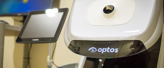
Optos Optomap Retinal Exam
|
The digital image is available on screen immediately for doctor and patient to review together, and will be saved in the patient's file to compare with future Optomap scans for signs of change.
In less than a second, the optomap is able to obtain an image of the retina for evaluation. The optomap retinal examination does not require the eyes to be dilated. |
Many people are uncomfortable with dilation because vision becomes blurry and sensitive to light for a period of time after the examination.
Humphrey Visual Field
|
The Humphrey visual field is a diagnostic test to measure visual fields, or perimetry. The Humphrey visual field test measures the entire area of peripheral vision that can be seen while the eye is focused on a central point. During this test, lights of varying intensities appear in different parts of the visual field while the patient's eye is focused on a certain spot. The perception of these lights is charted and then compared to results of a healthy eye at the same age of the patient to determine if any damage has occurred.
|
This procedure is performed in about 20 minutes and is effective in diagnosing and monitoring glaucoma. Patients with glaucoma will undergo this test on a regular basis to determine the progress of the disease. The Humphrey visual field test can also be used to detect conditions within the optic nerve of the eye, and certain neurological conditions as well.
OptoVue
iVue SD OCT
Conditions Detected With An OCT
The images can help with the detection and treatment of serious eye conditions such as:
OCT uses technology that is similar to that of a CT scan of internal organs. With the scattering of light it can rapidly scan the eye to create an accurate cross-section. Each layer of the retina can be evaluated and measured and compared to normal, healthy images of the retina.
The images can help with the detection and treatment of serious eye conditions such as:
- Macular hole
- Macular swelling
- Optic nerve damage
- Age-related macular degeneration
- Macular pucker
- Glaucoma
- Cataracts
- Diabetic eye disease
- Vitreous hemorrhage
OCT uses technology that is similar to that of a CT scan of internal organs. With the scattering of light it can rapidly scan the eye to create an accurate cross-section. Each layer of the retina can be evaluated and measured and compared to normal, healthy images of the retina.
iCare Tonometry
|
During your exam, our technicians will check your Intra Occular Pressure (IOP) with an Icare® tonometer. Based on a proven accurate measuring principle known as rebound technology, a light weight probe is used to make momentary and gentle contact with the cornea. The rebound technology eliminates corneal disruption and patient discomfort. This painless screening takes only a few seconds.
|
Medmont Corneal Topographer
|
The Medmont E300 offers extreme accuracy for the mapping of a patient's cornea. Used extensively by the top contact lens manufactures and optometry universities throughout the world, the E300 is considered the gold standard for fitting specialty contact lenses. With the ability to map out to 14mm, using composite mapping, it gives the practitioner a very high percentage of first fit success rates. This can be extremely helpful when fitting patients who wear Scleral or Ortho-K lenses, or patients who have had difficulty finding a comfortable fitting lens in the past. This instrument is also a diagnostic tool used to precisely analyze tear film surface quality for patients with dry eye.
|


